• Professional healthcare
  • Products & Services
  • Inspiration
  • Support & Contact
  • Shop
‌
‌
‌
‌
‌
‌
‌
‌
‌
‌
‌
‌
‌
‌
‌
‌
‌
‌
‌
‌
  • Consumer productsConsumer products
    • Home
    • Support
    • Product registration
    • My Philips
  • Healthcare professionalsHealthcare professionals
    • Home
    • Products
    • Services
    • Specialties
    • Solutions
  • Other business solutionsOther business solutions
    • Lighting
    • Automotive
    • Dictation Solutions
    • More
    • Hearing solutions
    • Display solutions
  • About usAbout us
    • Home
    • News
    • Investor Relations
    • Careers
    • More
    • Innovation
    • Environmental, Social and Governance
  • Contact and supportContact and support
    • Customer support
    • Professional healthcare support
    • Company contacts
  • Stay up-to-dateStay up-to-date
  • Select your country / languageGlobal / English
  • Privacy Notice
  • Terms of use
  • Cookie notice
© Koninklijke Philips N.V., 2004 - 2025. Kaikki oikeudet pidätetään.
‌
‌
‌
‌
‌
‌
‌
‌
‌
‌
‌
‌
‌
‌
‌
‌
‌
‌
‌
‌
  1. Diagnostic imaging
  2. Magnetic resonance imaging
  3. MRI software
  4. Multi Nuclei - Brain
Multi Nuclei - Brain
Seamless integration of Multi Nuclei
MRI software
‌
‌
‌
‌
‌
‌
‌
‌
‌

Multi Nuclei - Brain

Seamless integration of Multi Nuclei
MRI software
Adding multi-nuclei to your Philips 3.0T MR system opens a window of research into other nuclei, in search of metabolic and functional information. Thanks to a seamless integration onto the 3.0T platform, multi-nuclei imaging and spectroscopy become part of your daily clinical workflow. With the dual tuned head coils you can perform brain exams, including acquisition of proton and other nuclei (31P, 13C, 23Na), without switching coils. A full brain study, including both proton (1H) and sodium (23Na) imaging can be completed in 30 minutes1, all organized in one ExamCard. And a sodium (23Na) brain scan can be completed in less than 15 minutes2. Multi-nuclei imaging or spectroscopy can be run and reconstructed directly from the standard user interface. The nucleus is just a scan parameter like any other sequence parameter. Reconstruction and viewing of multi-nuclei images or spectra, as well as the process for sending the data to PACS is fully integrated, so workflow does not differ from proton imaging.

All Philips professional healthcare customers can open, track and manage equipment service tickets in our Customer Services Portal at no cost. Register for an account here.
For all support resources and contact information, visit our Support hub.
Seamless integration of Multi Nuclei imaging and spectroscopy
Workflow does not differ from proton imaging
Brain exams, including proton and other nuclei, without switching coils

Clinical image gallery

  • Multi Nuclei Brain exam 1H and 23Na
    Multi Nuclei Brain exam 1H and 23Na
  • Multi Nuclei Brain exam 1H and 13C
    Multi Nuclei Brain exam 1H and 13C
  • Multi Nuclei Brain exam 1H and 31P
    Multi Nuclei Brain exam 1H and 31P

Recommended products

  • MR 7700 Unmatched performance and precision
    MR 7700
    Experience breakthrough innovation in 3.0T imaging with the unique design of the Philips MR 7700 imaging system, enhanced with XP gradients and artificial intelligence (AI). The system is built to address a pressing need to deliver on the clinical expectations of today, and to facilitate the most demanding research programs. The MR 7700 provides high accuracy, power, and endurance to support confident diagnosis for every patient. It is the system of choice for highest quality diffusion imaging and advanced neuroscience. Extend your scanning capabilities with a fully integrated multi-nuclei imaging and spectroscopy solution to explore new clinical pathways without sacrificing clinical imaging workflow or wide-bore patient comfort. What’s more? The MR 7700 promises a great experience for both users and patients through the ease-of-use features of a well-designed clinical 3.0T scanner together with a no compromise workflow. Now scientists and clinicians alike can schedule without conflict.
  • Ingenia Elition 3.0T X A revolutionary breakthrough in diagnostic quality – and speed
    Ingenia Elition 3.0T X
    The Philips Ingenia Elition solution offers cutting-edge MR imaging techniques, while setting new standards for clinical research in 3.0T imaging based on gradient- and RF designs. The Ingenia Elition delivers on superb image quality, and performs MRI exams up to 50% faster¹. Fast overall exam-time is achieved by improving patient handling setup time at the bore with the touchless guided patient setup, combined with accelerations in both 2D- and 3D scanning. Furthermore, the Ingenia Elition offers an immersive audio-visual experience to calm patients and guide them through MR exams.
  • Multi Nuclei MR Seamless integration of multi-nuclei
    Multi Nuclei MR
    Extend your scanning capabilities with a fully integrated multi-nuclei imaging and spectroscopy solution to explore new clinical pathways without sacrificing clinical imaging workflow or wide-bore patient comfort. Multi-nuclei (MN) imaging and spectroscopy typically involves a different software version, cumbersome user interface, and a dedicated coil. And scan times tend to be quite long, which can disrupt day-to-day imaging throughput. To advance clinical insights in this promising area, Philips has made multi-nuclei imaging and spectroscopy become part of your daily clinical workflow. 
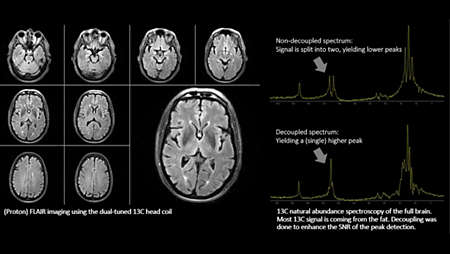
Adding multi-nuclei to your 3.0T MR system opens a window of research into other nuclei, in search of metabolic and functional information. It allows you to perform clinical imaging, spectroscopy and research studies of six different nuclei (1H, 31P, 13C, 23Na, 19F* and 129Xe*). Simply put, our multi-nuclei solution can be used across all anatomies.